Digital radiography in 3D
The device used for the radiography
The camera used for panoramic and cephalometric x-rays is different from the one used for routine x-rays. In order to photograph the entire mouth, the joints and the position of your teeth, the camera will rotate around the head and be oriented on your cheek or the front of your mouth.
Regular inspection of radiology equipment
X-ray equipment is inspected regularly. The dose of x-rays directed at your mouth is calibrated to minimize the risks associated with radiation exposure.

Why 3D radiography?
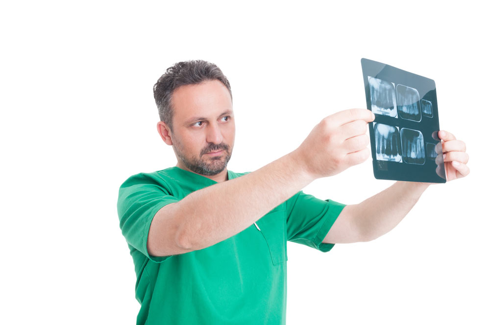
The advent of 3D digital radiography has greatly facilitated the work of specialists, in addition to reducing the radiation dose for the patient. The most commonly used 3D radiograph in implantology is the panoramic radiograph, which shows the teeth and bone structures of the patient’s maxillofacial area.
In order to plan the installation of dental implants, it is often necessary to obtain additional information that is unfortunately not available with the traditional 2D radiograph:
- the quantity and density of the alveolar bone in all three dimensions (height, width and depth);
- the position and anatomy of the maxillary sinuses for implants in the upper jaw;
- the precise position of the inferior alveolar nerve for implants in the lower jaw.
These three elements must absolutely be considered in implantology because of the complications that can occur during implant surgery. 3D digital radiography, also called three-dimensional imaging by cone-beam volumetric computed tomography (CBCT)(cone beam computed tomography (CBCT), is available in our office for surgical case planning.
Advantages of 3D digital radiography
Three-dimensional radiography offers several advantages for both the dentist and the patient.
For the patient:
- Less exposure to radiation (due to the technology used and the ability to take images in less time).
- Ease of understanding the specialist’s explanations (by the clarity and precision of the radiographic images). Indeed, several views can be displayed, allowing the specialist and the patient to see the structures from different angles.
For the dentist:
-
- The quality and quantity of information provided by TVFC on different types of tissues and organs, such as soft tissue, bones, muscles, nerves and even blood vessels, make the more accurate, timely and predictable implant installation planning. The specialist can obtain images of impacted teeth, the relationship of the teeth to each other, the quality and volume of the jawbone, the maxillary sinuses and the inferior alveolar nerve, which are not available with traditional 2D radiographs.
- The specialist knows exactly the ideal position for each implant,to a fraction of a millimeter, which facilitates surgery and allows it to achieve unparalleled precision.
- Damage to neighbouring structures to the implants (remaining teeth, inferior alveolar nerve and maxillary sinuses),can be minimized.
- A single 3D radiograph can generate hundreds of images
When the X-ray machine’s sensors finish acquiring the data, it is sent to a computer with software to produce the various views. A single three-dimensional X-ray can generate hundreds of images and even 3D videos of the patient’s face.
- 3D digital radiography: sharper images
The main innovations of 3D scans are the absence of distortion and the high resolution obtained by the powerful data reconstruction software. A single data acquisition by a TVFC allows to create several views that can be manipulated in all directions by the dentist
When the X-ray machine’s sensors finish acquiring the data, it is sent to a computer with software to produce the various views. A single three-dimensional X-ray can generate hundreds of images and even 3D videos of the patient’s face.
- 3D digital radiography: sharper images
The main innovations of 3D scans are the absence of distortion and the high resolution obtained by the powerful data reconstruction software. A single data acquisition by a TVFC allows to create several views that can be manipulated in all directions by the dentist
